NeuroCSF: an fMRI method to measure contrast sensitivity function in human visual cortex
Goulet L, Farivar R.
,
J Neurophysiol
,
2025
@article{goulet2025neurocsf,
title={NeuroCSF: an fMRI method to measure contrast sensitivity function in human visual cortex},
author={Goulet, Laurie and Farivar, Reza},
journal={Journal of Neurophysiology},
volume={133},
pages={1699–1716},
year={2025},
publisher={American Physiological Society},
doi={10.1152/jn.00463.2024}
}
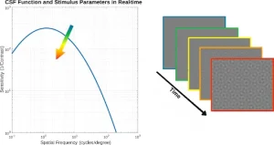
The contrast sensitivity function (CSF) describes a range of spatial frequencies (SFs) that are detectable at a given level of contrast and is a very valuable tool both in clinical and fundamental research. However, despite its immense value, the full potential of the CSF has not been utilized in every aspect of clinical research due to time limits and patient factors. We propose neuroCSF as a new method for measuring the CSF across the visual field directly from brain activity and with minimal demand from participants. NeuroCSF is a computational model that estimates voxel-wise CSF parameters (i.e., peak contrast sensitivity, peak spatial frequency, and spatial frequency bandwidth) from functional magnetic resonance imaging (fMRI) signals under controlled visual stimulation conditions. We observe that across early visual areas (V1, V2, and V3), the CSF peak spatial frequency and spatial frequency cutoff are significantly higher for foveal eccentricity and decrease at parafoveal eccentricities. Conversely, SF bandwidth slowly increases with eccentricity, while peak contrast sensitivity remains constant with eccentricity for all early visual areas. The neuroCSF approach opens new perspectives for the study of cortical visual functions in various disorders where the CSF is impacted, such as amblyopia, traumatic brain injury, and multiple sclerosis.
Plain Language Summary